
Enhancing Microscopy with SWIR Cameras
Microscopy is a fundamental tool in scientific research, allowing scientists to observe and analyze the minutiae of biological specimens, materials, and devices. While visible light microscopy provides valuable insights, the integration of shortwave infrared (SWIR) cameras introduces a new dimension to imaging. SWIR technology, particularly through Princeton Infrared Technologies' MVCam SWIR camera, enhances microscopy capabilities and addresses the limitations of conventional imaging methods.
Shortwave infrared (SWIR) imaging captures light in the wavelength range of approximately 0.4 to 1.7 µm. Unlike visible light, SWIR can penetrate deeper into materials and tissues, providing unique contrasts and details that are not visible with traditional visible light microscopy.
The advantages of SWIR in microscopy include enhanced contrast and detail, improved penetration and reduced scatter and noise. SWIR imaging provides superior contrast and detail for distinguishing between materials and structures that are difficult to differentiate using visible light. SWIR light can penetrate deeper into biological tissues and materials, allowing for imaging of internal structures that are opaque to visible light. SWIR wavelengths are less scattered by particles and imperfections, leading to clearer images in challenging conditions.
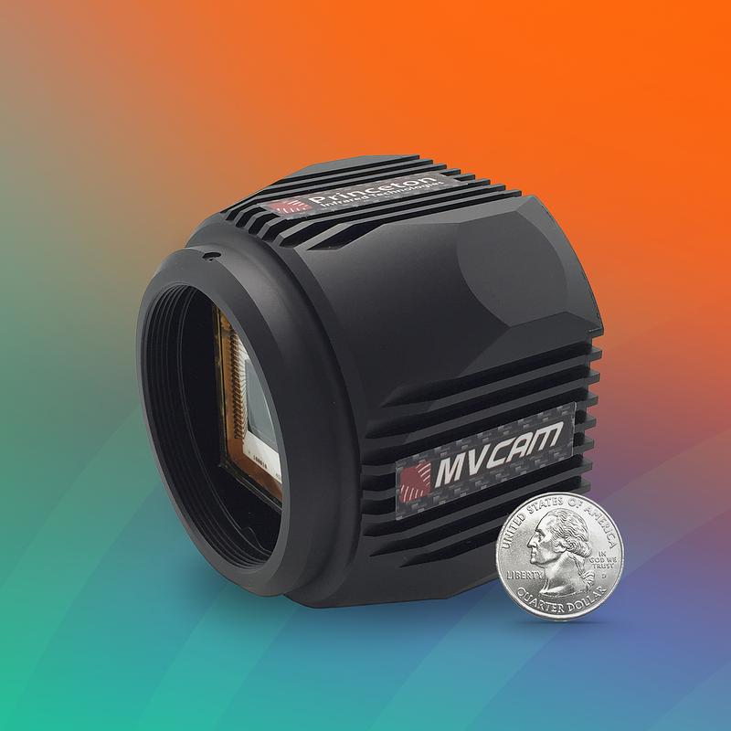
The MVCam SWIR camera represents the latest advancements in SWIR imaging technology, specifically designed to enhance microscopy applications. Key features of the MVCam include high resolution (1280x1024 megapixels), fast frame rate (up to 90 frames per second), InGaAs sensor technology with a 12µm pixel pitch, 0.4-1.7µm wavelength range covering both visible and SWIR for versatile imaging. The MVCam also has an integrated single-stage thermoelectric cooler for consistent sensor temperature, minimizing thermal noise and medium and base Camera Link™ configurations for flexible data output and system integration.
The microscopy applications for the MVCam include biological research and material science.
SWIR imaging with the MVCam can significantly advance biological research by providing deeper insights into cellular and tissue structures by observing cellular processes and structures with enhanced contrast and detail. The MVCam is also able to visualize internal tissue structures in tissue analysis and study tissue morphology with improved penetration and reduced scattering.
In material science, the MVCam SWIR cameras offer valuable advantages for analyzing materials and detecting defects. In material characterization, the MVCam can examine material surfaces and internal structures with high resolution, revealing hidden defects and characteristics. The MVCam can also assist with quality control by monitoring and inspecting materials in real-time, ensuring consistent quality and performance.
The MVCam SWIR Camera’s Camera Link™ interface ensures compatibility with a wide range of microscopy systems, allowing for seamless integration. The flexible data output options accommodate different frame rates and resolutions, catering to various application needs. To maximize the benefits of SWIR imaging, it is essential to optimize camera settings and integrate the SWIR camera effectively into your microscopy system.
The integration of SWIR cameras, such as the Princeton Infrared Technologies MVCam SWIR camera, represents a significant advancement in microscopy. By extending imaging capabilities beyond the visible spectrum, SWIR technology provides enhanced contrast, detail, and penetration, opening new possibilities for research and analysis across various fields.

 MVCam
MVCam